• iconNews
  • videos
  • entertainment
  • Home
  • News
    • UK News
    • US News
    • Australia
    • Ireland
    • World News
    • Weird News
    • Viral News
    • Sport
    • Technology
    • Science
    • True Crime
    • Travel
  • Entertainment
    • Celebrity
    • TV & Film
    • Netflix
    • Music
    • Gaming
    • TikTok
  • LAD Originals
    • Say Maaate to a Mate
    • Daily Ladness
    • Lad Files
    • UOKM8?
    • FreeToBe
    • Extinct
    • Citizen Reef
  • Advertise
  • Terms
  • Privacy & Cookies
  • LADbible Group
  • UNILAD
  • SPORTbible
  • GAMINGbible
  • Tyla
  • UNILAD Tech
  • FOODbible
  • License Our Content
  • About Us & Contact
  • Jobs
  • Latest
  • Topics A-Z
  • Authors
Facebook
Instagram
X
Threads
Snapchat
TikTok
YouTube

LAD Entertainment

YouTube

LAD Stories

Submit Your Content
First ever 'Google Earth' organ video shows inside healthy and diseased hearts in extraordinary detail

Home> News> Science

Published 11:46 17 Jul 2024 GMT+1

First ever 'Google Earth' organ video shows inside healthy and diseased hearts in extraordinary detail

Instead of stalking your neighbour's garden, now you can look at a stranger's heart

Jess Battison

Jess Battison

Look, I don’t know who I even need to say this to, but as much as you might like to think it does, a heart doesn’t look anything like the ones we’d scribble onto our mate’s planners at school.

Obviously not. But still, it’s not exactly often that we get to have a deep, good old look at a real human heart.

And now, the first ever ‘Google Earth’ organ video shows us the insides of healthy and diseased hearts in extraordinary detail.

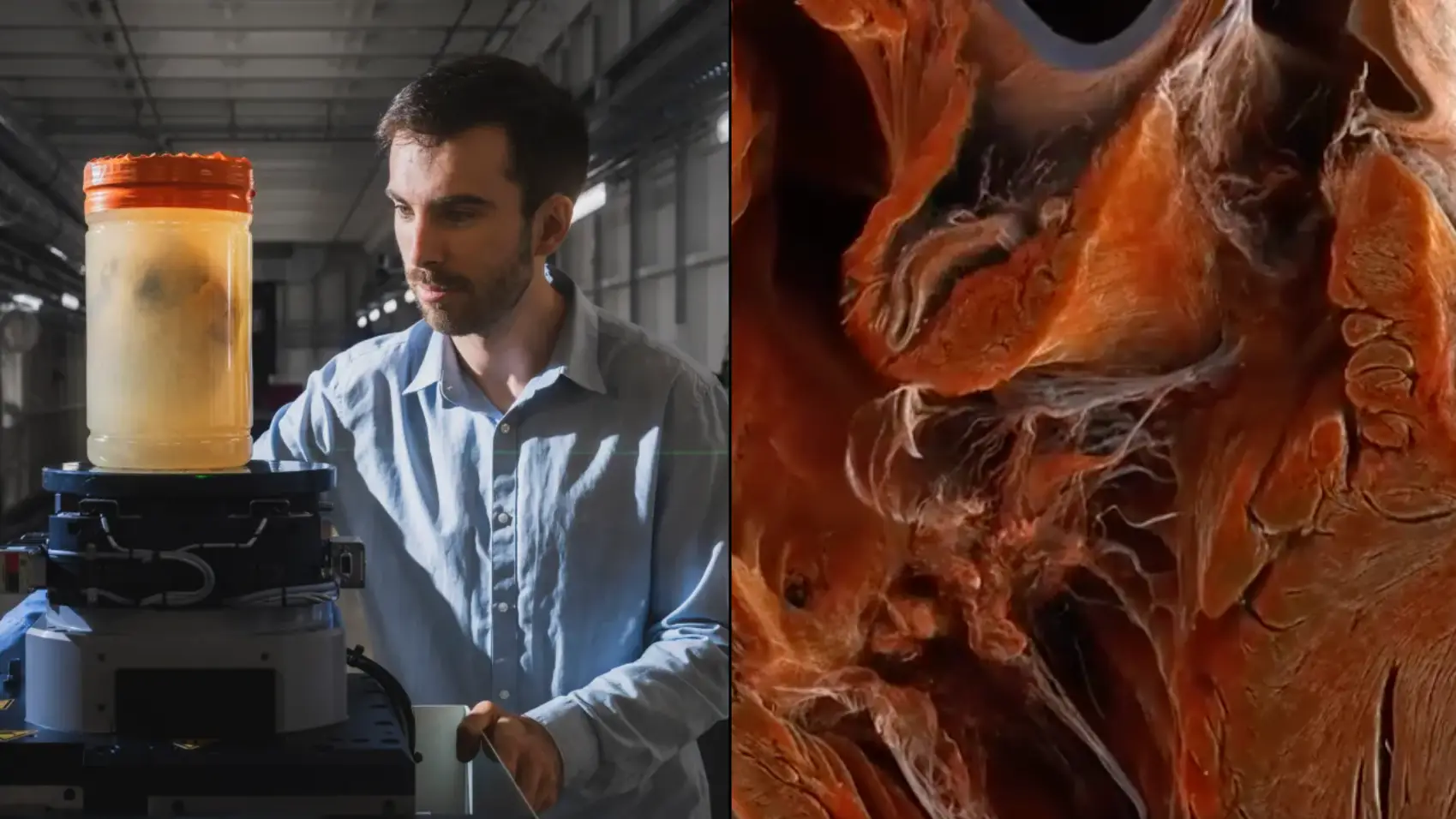
Experts at University College London (UCL) and the European Synchrotron Radiation Facility (ESRF) used a new X-ray technique to capture the anatomical structure of them right down to a teeny-tiny 20 micrometres.

Advert

So, while you might spend your free time stalking random streets on Google Earth – hey, no judgement – that’s pretty much the detail you can explore a heart to with this.

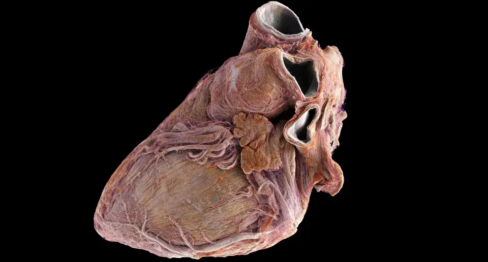
The video compares two whole hearts from two adult donors. And while you can clearly see a defined shape of the healthy heart, the diseased one appears rounder with more withered vessels and muscle fibres.

“The atlas that we’ve created in this study is like having Google Earth for the human heart,” explained Professor Peter Lee, senior author of the study from UCL Mechanical Engineering.

“It allows us to view the whole organ at global scale, then zoom in to street level to look at cardiovascular features in unprecedented detail.”

The experts scanned two different human hearts. (ESRF/Stef Candé)

Advert

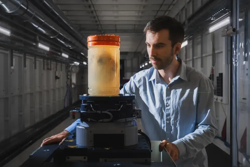
At France’s ESRF, the team used the X-ray technique hierarchical phase-contrast tomography (HiP-CT) to image the hearts down to this minuscule scale.

“One of the major advantages of this technique is that it achieves a full 3D view of the organ that’s around 25 times better than a clinical CT scanner,” Lee added.

READ MORE:

YOUTUBER WARNS OF MEAL THAT KILLED HIS GIRLFRIEND

SUICIDE POD INVENTOR WANTS TO INSTALL THEM IN UK

Advert

“In addition, it can zoom in to cellular level in selected areas, which is 250 times better, to achieve the same detail as we would through a microscope but without cutting the sample.

“Being able to image whole organs like this reveals details and connections that were previously unknown.”

They got a good look at a healthy heart. (UCL/ESRF)

The reason the researches used hearts from deceased donors is simply because it just wouldn’t be possible to image a living heart in this way as the radiation dose would be far too high for it to be safe.

Experts hope these new images will help with increasing the understanding of cardiovascular disease.

Advert

“We now have a way to determine differences in the thickness of tissue and fat layers located between the outer surface of the heart and the protective sac surrounding the heart, which could be relevant when treating arrhythmia,” said Professor Andrew Cook, heart anatomist from the UCL's cardiovascular science institute.

The study was published yesterday on Radiology.

Featured Image Credit: UCL/ESRF/Stef Candé

Topics: Health, Science, Technology

Jess Battison
Jess Battison

Jess is a Senior Journalist with a love of all things pop culture. Her main interests include asking everyone in the office what they're having for tea, waiting for a new series of The Traitors and losing her voice at a Beyoncé concert. She graduated with a first in Journalism from City, University of London in 2021.

X

@jessbattison_

Advert

Advert

Advert

  • Moment of death was recorded for first ever time and reveals what our final thoughts could be
  • 'Most extraordinary instrument ever built by humanity' could unravel secrets of the universe
  • Fascinating video shows how fasting works inside the human body
  • Billionaire completes 'dangerous' first-ever private spacewalk as first words are revealed

Choose your content:

3 hours ago
4 hours ago
  • 3 hours ago

    World's 'first flying car' is going on sale much sooner than you think

    Flying cars are still something for the future, but apparently the rapidly approaching future

    News
  • 4 hours ago

    Experts issued warning over certain tattoo colour that could increase risk of deadly disease

    There can be some long-term health risks to going under the tattoo needle

    News
  • 4 hours ago

    Man who didn't sleep for a record 264 hours suffered from horrendous and potentially deadly side effects

    He smashed the record but suffered dangerous side effects in the process

    News
  • 4 hours ago

    The targets Iran could strike as it issues chilling threat to UK amid ongoing conflict

    The world isn't feeling particularly safe right now

    News